
Eos DR (DDDR)


Eos D (DDD)
THE EVOLUTION of hemodynamic pacingwith innovative diagnostic applications
 Advanced diagnostic description of tachyarrhythmic episodes, with simultaneous TVI and iECG acquisition
The cyclic fluctuation of Transvalvular Impedance (TVI) reflects the mechanical activity of the ventricle
The integrated intracardiac electrogram (iECG) allows an accurate evaluation of the electrical activity
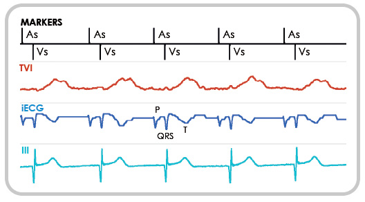 Atrial and ventricular electric signals are displayed in a single tracing, featuring properties similar to the surface ECG. As in the case of the ECG, the ventricular activation pattern affects the signal waveform. The reported example shows the difference in iECG morphology in case of ventricular sensing
or pacing.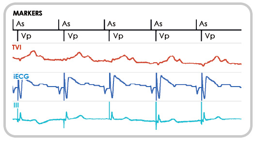
The morphological evaluation provides a quick and precise discrimination of supraventricular and ventricular arrythmias. iECG and TVI are automatically stored in memory during a tachycardia. At the follow-up check, the two tracings can be simultaneously transmitted by realtime telemetry, even during a threshold test.
Ventricular pacing reduction with hemodynamic surveillance relying on the TVI signal
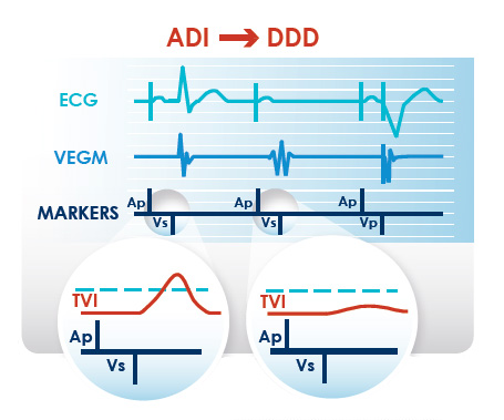 The ejection surveillance based on TVI immediately enables dual-chamber pacing in case of false inhibition of ventricular stimulation.
The maximum PR interval allowed is dynamically shortened according to the cardiac rate.
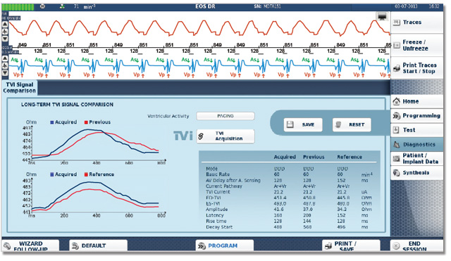
Long-term comparison of ventricular mechanics
![]() TVI waveforms recorded at different times are compared to assess the patient’s emodynamic stability.
TVI waveforms recorded at different times are compared to assess the patient’s emodynamic stability.
![]() The main waveform parameters are measured automatically.
The main waveform parameters are measured automatically.
![]() Trends of the daily average of diastolic and systolic TVI are stored in memory between two follow-up checks.
Trends of the daily average of diastolic and systolic TVI are stored in memory between two follow-up checks.
iECG allows the discrimination of ventricular and supraventricular tachycardias
according to the morphology of the QRS complex
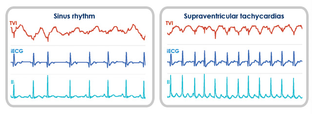
In the event of SVT, the morphology of the ventricular component of the iECG is unaffected. The presence of regular TVI fluctuation indicates a preserved pump function.
TVI provides information on the presence of hemodynamic activity and the stability of the pump function
In case of VT, the iECG features wide complexes, easy to distinguish from the signal recorded in the presence of AV conduction.
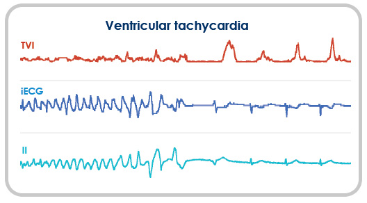
The example shows a spontaneously terminated VT. The TVI tracing reveals a marked depression of the pump function during the tachycardia, followed by the prompt recovery of systolic ejection when the rhythm is re-established.
![]() For more information or to get technical support on our products please CONTACT US >>
For more information or to get technical support on our products please CONTACT US >>
Products | Technology | Support | Contacts